| | |
|
| |
| | |
|
|
J. MORITA Veraviewepocs 3D R100
J. MORITA Veraviewepocs 3D R100
3D, Panoramic and Cephalometric X-Ray
Downloads
Featuring Morita's patent pending 3D Reuleaux Full Arch Field of View
Veraviewepocs 3D R100 is the latest addition to J. Morita’s 3D product line. This unit’s completely unique 3D Reuleaux Full Arch FOV (field of view) abandons the typical cylinder with a new convex triangle shape. By more closely matching the natural dental arch form, this groundbreaking FOV reduces dosage by excluding areas outside the region of interest and allows a complete scan of the maxilla and/or the mandible.
High resolution, 125 µm voxel images provide a clear observation of the periodontal pocket, the periodontal membrane, and the alveolar bone, as well as soft tissue such as the maxillary sinus membrane. A special dose reduction mode lowers exposure for soft tissue which reduces dosage to a mere 60% of the standard mode.
Multifunctional, it also offers panoramic and cephalometric imaging. With a total of 6 fields of view, 3D R100 is ideal for implantology as well as being suitable for periodontology, orthodontics, endodontics, oral surgery, and general dentistry.
Learn more about Morita 3D technology in scientific research.
FEATURES
- 3D, panoramic, and cephalometric capabilities
- New 3D Reuleaux Full Arch FOV offers a full arch scan
- New Dose Reduction Feature lowers X-ray exposure 30% to 40%
- Three options for easy and accurate 3D positioning
- Seven pre-programmed panoramic functions with magnification options
- New panoramic image layer adjustment options
- Super high resolution: 125 µm (micrometer) fine voxel
- 7.4 second panoramic; 9.4 second 3D scan
- Six FOV: Ø 40 x H 40 mm, Ø 40 x H 80 mm, Ø 80 x H 50 mm, Ø 80 x H 80 mm,
3D Reuleaux Full Arch FOV: Ø 100 mm x H 50 mm, Ø 100 mm x H 80 mm
Downloads
3D R100 offers a total of 6 exposure areas from 40 x 40 mm up to 100 x 80 mm for various diagnostic needs. The new full arch scan captures the maxilla and/or the mandible with the equivalent of 100 mm in diameter and two height options of 50 or 80 mm. Its full arch capability, reduced dosage, and exceptional clarity are ideal features for implant planning and oral surgery.
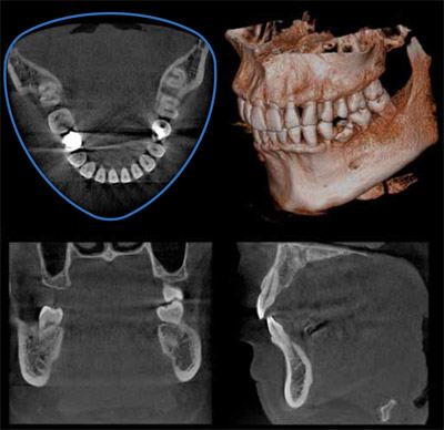
Successful placement of implants starts with the very critical and detailed planning process. Identification of structures such as the sinus cavity, inferior alveolar nerve, and clear views of the bone structure are needed. Veraviewepocs 3D R100 is ideal for implant planning with full arch imaging, industry leading clarity, low dosage to the patient and advanced implant planning software features. By converting images to DICOM formats, implant simulation can also be performed with other third party software. Oral surgery 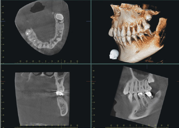 The patient presented with pain in the maxillary left region. A cone beam CT image was taken with the 3D R100 and it was revealed that tooth #16 was in fact impacted and was causing problems for tooth #15. The axial view demonstrated extensive bone loss near the apical area of #15 due to the lack of arch space needed for #16 to erupt. The coronal view showed bone destruction all the way through the furcation of #15. The sagittal image not only shows the loss of osseous support around the entire apex of #15, but also shows damage to the sinus floor and mucosal thickening. The patient presented with pain in the maxillary left region. A cone beam CT image was taken with the 3D R100 and it was revealed that tooth #16 was in fact impacted and was causing problems for tooth #15. The axial view demonstrated extensive bone loss near the apical area of #15 due to the lack of arch space needed for #16 to erupt. The coronal view showed bone destruction all the way through the furcation of #15. The sagittal image not only shows the loss of osseous support around the entire apex of #15, but also shows damage to the sinus floor and mucosal thickening.
Endodontics 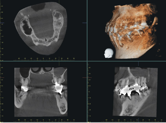 The patient presented with a radiolucency around tooth #14 that had previously been treated endodontically. Conventional 2D imaging was inconclusive so a cone beam CT image was taken with the 3D R100. The sagittal and coronal views both showed that the endodontic therapy was failing and there were apical lesions on both the buccal and palatal roots. The sagittal view clearly confirms a perforation of the Schneiderian membrane, while the coronal view identifies odontogenic maxillary sinusitis and mucosal thickening. The damage to the sinus membrane may have been overlooked if this case was diagnosed and treatment planned with an image that did not show the problem so clearly. The patient presented with a radiolucency around tooth #14 that had previously been treated endodontically. Conventional 2D imaging was inconclusive so a cone beam CT image was taken with the 3D R100. The sagittal and coronal views both showed that the endodontic therapy was failing and there were apical lesions on both the buccal and palatal roots. The sagittal view clearly confirms a perforation of the Schneiderian membrane, while the coronal view identifies odontogenic maxillary sinusitis and mucosal thickening. The damage to the sinus membrane may have been overlooked if this case was diagnosed and treatment planned with an image that did not show the problem so clearly.
Implantology
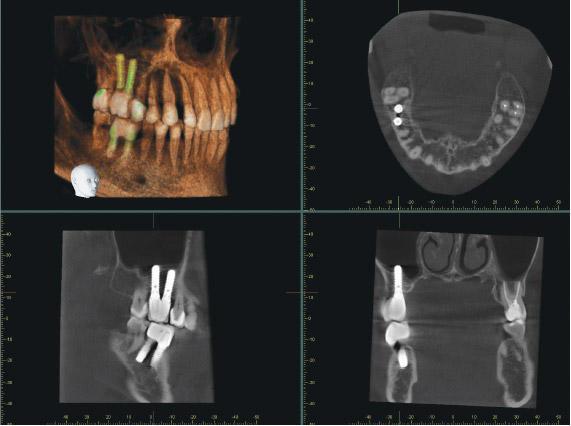 The patient was seen for a routine follow up visit following implant placement of tooth #3. The implant had been placed 9 years earlier. The coronal, sagittal, and axial views all confirm good quality bone around the implant and an absence of any pathology related to the treatment. The fine detail of the bone, especially in the coronal view, allows the clinician and the patient to feel confident about the current health of the implant. The patient was seen for a routine follow up visit following implant placement of tooth #3. The implant had been placed 9 years earlier. The coronal, sagittal, and axial views all confirm good quality bone around the implant and an absence of any pathology related to the treatment. The fine detail of the bone, especially in the coronal view, allows the clinician and the patient to feel confident about the current health of the implant.
|
|